


Rotationally Resliced 3D Prostate Segmentation of MR images Using Bhattacharyya Similarity and Active Band Theory
Authors: Tang, Zhixian; Wang, Manning; Song, Zhijian
Physica Medica (2018)
Purpose: In this article, we propose a novel, semi-automatic segmentation method to process 3D MR images of the prostate using the Bhattacharyya coefficient and active band theory with the goal of providing technical support for computer-aided diagnosis and surgery of the prostate.
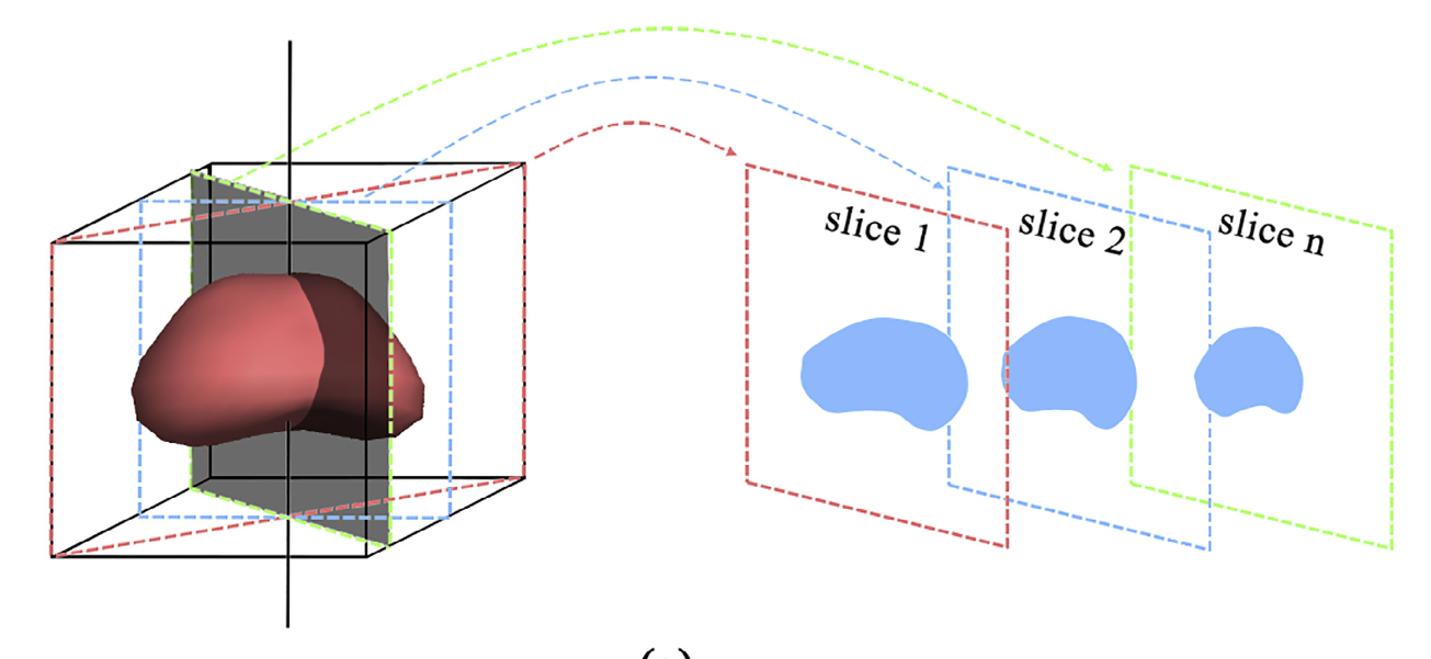
Methods: Our method consecutively segments a stack of rotationally resectioned 2D slices of a prostate MR image by assessing the similarity of the shape and intensity distribution in neighboring slices. 2D segmentation is first performed on an initial slice by manually selecting several points on the prostate boundary, after which the segmentation results are propagated consecutively to neighboring slices. A framework of iterative graph cuts is used to optimize the energy function, which contains a global term for the Bhattacharyya coefficient with the help of an auxiliary function. Our method does not require previously segmented data for training or for building statistical models, and manual intervention can be applied flexibly and intuitively, indicating the potential utility of this method in the clinic.
Results: We tested our method on 3D T2-weighted MR images from the ISBI dataset and PROMISE12 dataset of 129 patients, and the Dice similarity coefficients were 90.34 ± 2.21% and 89.32 ± 3.08%, respectively. The comparison was performed with several state-of-the-art methods, and the results demonstrate that the proposed method is robust and accurate, achieving similar or higher accuracy than other methods without requiring training.
Conclusion: The proposed algorithm for segmenting 3D MR images of the prostate is accurate, robust, and readily applicable to a clinical environment for computer-aided surgery or diagnosis.