

Multi-Label Conditioned Diffusion for Cardiac MR Image Augmentation and Segmentation
Bioengineering (IF=3.7)
Abstract
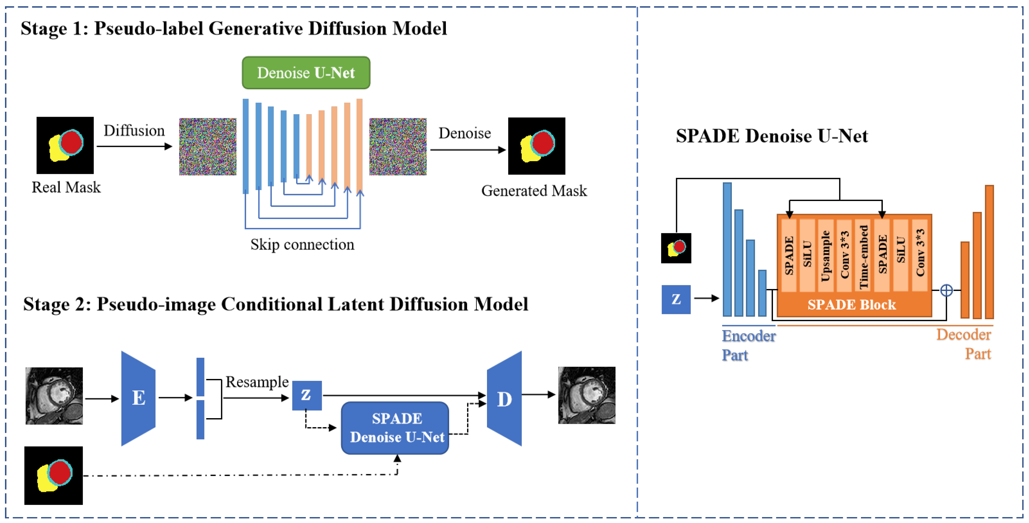
Accurate segmentation of cardiac MR images using deep neural networks is crucial for cardiac disease diagnosis and treatment planning, as it provides quantitative insights into heart anatomy and function. However, achieving high segmentation accuracy relies heavily on extensive, precisely annotated datasets, which are costly and time-consuming to obtain. This study addresses this challenge by proposing a novel data augmentation framework based on a condition-guided diffusion generative model, controlled by multiple cardiac labels. The framework aims to expand annotated cardiac MR datasets and significantly improve the performance of downstream cardiac segmentation tasks. The proposed generative data augmentation framework operates in two stages. First, a Label Diffusion Module is trained to unconditionally generate realistic multi-category spatial masks (encompassing regions such as the left ventricle, interventricular septum, and right ventricle) conforming to anatomical prior probabilities derived from noise. Second, cardiac MR images are generated conditioned on these semantic masks, ensuring a precise one-to-one mapping between synthetic labels and images through the integration of a spatially-adaptive normalization (SPADE) module for structural constraint during conditional model training. The effectiveness of this augmentation strategy is demonstrated using the U-Net model for segmentation on the enhanced 2D cardiac image dataset derived from the M&M Challenge. Results indicate that the proposed method effectively increases dataset sample numbers and significantly improves cardiac segmentation accuracy, achieving a 5% to 10% higher Dice Similarity Coefficient (DSC) compared to traditional data augmentation methods. Experiments further reveal a strong correlation between image generation quality and augmentation effectiveness. This framework offers a robust solution for data scarcity in cardiac image analysis, directly benefiting clinical applications.